Your Guide to Understanding an Ultrasound Scan

Author: Nnenna Chiloli, MBBS, MPH
Medical Reviewer: Oke Ikpekpe, MSc (PH)
Highlights
- Ultrasound uses high-frequency sound waves to create real-time images of organs, tissues, and blood flow.
- Ultrasound can help diagnose and monitor conditions affecting the abdomen, pelvis, heart, and blood vessels.
- Ultrasound is widely used in pregnancy to monitor fetal growth and assess the position of the placenta.
- Proper preparation for an ultrasound depends on the body area being examined.
- Ultrasound is generally painless and does not use ionising radiation.
- Although ultrasound is considered safe, trained professionals should use appropriate settings and the lowest settings needed to obtain clear diagnostic images.
Introduction
An ultrasound scan, also known as sonography, is a medical test that uses high-frequency sound waves to create live images of the inside of the body [1]. Unlike X–rays, ultrasound does not use ionising radiation, making it particularly useful during pregnancy.
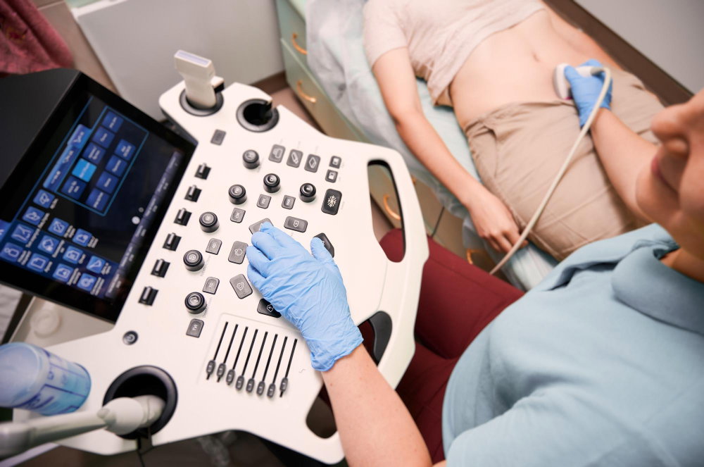
It works by emitting sound waves from a handheld device called a transducer (or probe). These waves travel into the body and bounce off internal structures like organs and tissues.
The probe receives the returning echoes, which are processed by a computer to produce real-time images on a monitor. These images allow healthcare professionals to see the size, shape, and movement of internal organs. Doppler ultrasound is used to assess blood flow.
In this article, you will learn what an ultrasound scan is, why it is done, what conditions it can help diagnose, and its benefits, limitations, and possible risks.
Why Is an Ultrasound Scan Done?
Ultrasound scans are performed for several reasons, such as:
Diagnostic purposes
Doctors use ultrasound to investigate symptoms like pain, swelling, or infection. They help identify the possible cause of symptoms by providing clear images of soft tissues that may not show up well on other imaging tests. For example, it can help locate a lump in the breast.
Monitoring existing conditions
For patients with known health issues, ultrasound is used to monitor changes in a condition or assess the response to treatment. This is particularly common in obstetrics, where it monitors the growth and health of a developing fetus throughout pregnancy.
Guiding medical procedures
Ultrasound provides real-time guidance for doctors during some procedures.It is used to:
- Guide needles for biopsies (taking tissue samples)
- Assist in fluid drainage
- Help place catheters in blood vessels [2].
The guidance from ultrasound results can help healthcare professionals perform these procedures more accurately and reduce certain risks.
What Conditions Can an Ultrasound Scan Help Diagnose?
Ultrasound helps in the diagnosis of a variety of health conditions such as:
Abdominal and Pelvic Health
Ultrasound is a primary tool for examining organs in the abdomen, including the liver, gallbladder, kidneys, pancreas, and spleen. It can:
- Detect gallstones and kidney stones
- Assess liver size, texture, and features suggestive of chronic liver disease
- Detect cysts and abnormal masses in the abdomen.
In the pelvis, it is used to investigate issues with the bladder, uterus, and ovaries, such as fibroids, ovarian cysts, or causes of abnormal bleeding.
Heart health
Echocardiography is a specialised ultrasound of the heart used to evaluate heart structures and function. It can diagnose:
- Heart valve abnormalities or regurgitation.
- Abnormalities in the heart's structure and the size of its chambers.
- Pericardial effusion (fluid around the heart).
Vascular ultrasound/ Doppler assesses blood flow through arteries and veins and can help detect:
- Blood clots such as deep vein thrombosis
- Narrowing or blockage of vessels
Abnormal blood flow
Pregnancy Health
Ultrasound is the standard for monitoring pregnancy. It helps determine:
- Pregnancy dating
- Number of babies
- Abnormalities in the baby
- Placental position and fetal growth
Emergency and Trauma
In emergencies, scans such as focused assessment with sonography for trauma (FAST) are used to quickly check for life-threatening conditions like internal bleeding or air around a collapsed lung (pneumothorax) [3].
Breast Health
Depending on the age, symptoms, and clinical circumstances, ultrasound may be used to:
- Evaluate a palpable lump
- Distinguish cystic from solid lesions
- As a supplementary test after mammography.
Breast ultrasound does not replace mammography, and any suspicious finding may require a biopsy.
Musculoskeletal
Ultrasound can assess:
- Muscle injury [4]
- Tendon tears
- Excess fluid in the joints
- How tendons move during examination
What Are The Limitations of an Ultrasound Scan?
Limitations to ultrasound include the following:
- Gas in the bowel can make it difficult to obtain clear images.
- Ultrasound cannot see clearly through bone, making it difficult to examine structures behind it.
- Scan quality also depends on the skill and experience of the person performing it.
- Obesity can reduce the image quality
- A normal ultrasound does not always rule out disease.
- Computerized Tomography (CT) or Magnetic Resonance Imaging (MRI) can better assess some abnormalities.
What Types of Ultrasound Scans Are Available?
Different types of ultrasound technology are used depending on the medical question being answered.
External Ultrasound
This is the most common type. The probe is moved over the skin of the area being examined, such as the abdomen or neck. A gel is applied to help the sound waves travel between the probe and the skin
Internal Ultrasound
Some scans require the probe to be placed inside the body for a clearer view of certain organs. Examples are:
A small covered probe is inserted into the vagina to view the uterus and ovaries
A small, covered probe is inserted into the rectum to examine the prostate gland.
- Doppler Ultrasound
This measures blood flow through your blood vessels.
How to Prepare for an Ultrasound Scan
Preparation depends on the area being scanned. Your clinic will provide specific instructions. However, general preparation includes the following:
Clothing
Wear loose, comfortable clothing. You may be asked to remove jewelry or change into a hospital gown.
Medication
You can usually take your regular medicines, but check with your healthcare provider if you have been asked to fast or have been given different instructions. Specific instructions include:
Abdominal or belly Scans
Follow the specific preparation instructions given by the imaging center. You may need to fast before a gallbladder scan, while an ultrasound of the pelvis or bladder may require you to drink water and keep your bladder full.
Breast Ultrasound
Usually requires no special preparation.
For an internal ultrasound, a healthcare professional gently inserts a covered, lubricated probe into the vagina or rectum. They will explain the procedure and obtain your consent before starting. You can ask them to stop if you feel uncomfortable.
What Happens During an Ultrasound Scan?
Understanding the process can help you feel more at ease during your appointment.
Positioning
You will be asked to lie on a comfortable examination couch. Depending on the scan, you may be asked to lie on your back or side.
Application of gel
The healthcare provider will apply a clear, water-based gel to your skin over the area to be examined. This gel may feel cold, but it helps the sound waves travel between the probe and your body.
Scanning
The healthcare professional will gently press the probe against your skin and move it back and forth. They will watch the images on a computer screen and may capture still photos or video clips.
Adjustments
The lights in the room may be dimmed to make the screen easier to see. You might be asked to hold your breath for a few seconds or change positions to get a better view.
Duration
A typical scan takes 10 to 45 minutes, though more complex examinations can take longer.
After the Procedure
Once the scan is finished, the gel will be wiped off your skin. You can dress immediately and resume your normal activities. If you have had an additional procedure, your healthcare provider will explain any special instructions.
Is an Ultrasound Scan Painful?
For most patients, an ultrasound is a painless and comfortable experience. A routine external diagnostic ultrasound does not involve needles or a cut in the body. You cannot feel the sound waves themselves; however, you may feel some pressure as the sonographer presses the probe against your skin to get the best possible image. If the area being scanned is tender, this pressure might be slightly uncomfortable.
Understanding Ultrasound Scan Results
The images produced during the scan are interpreted by a specialist.The specialist will write a report based on their findings. This report is sent to the doctor who referred you for the scan. The sonographer may sometimes give you a general idea of what they saw, but your referring doctor is the one who will discuss the full results and any necessary treatment plans with you.
Some results may be available immediately, while others take longer. Before leaving, ask when and how you will receive your results. If the scan shows an abnormality or does not provide enough information, your doctor may recommend another ultrasound, further imaging or other tests.
What Are the Benefits of an Ultrasound Scan?
Ultrasound offers several advantages over other types of medical imaging.
Safety
It does not use ionizing radiation, which eliminates the risks associated with X-rays.
Non-invasive
It provides a clear view of internal organs without the need for surgery or injections.
Realtime Images
It shows real-time images of organs and blood flow as it happens, which is vital for heart and fetal monitoring.
Accessibility
Ultrasound machines are often portable and can be used at the patient's bedside.
Cost-effective
It is often less expensive than CT or MRI scans.
Are There Any Risks or Side Effects of Ultrasound?
Ultrasound is generally considered very safe when medically necessary and performed appropriately by trained professionals [5]. However, unnecessary and/or prolonged exposure should be avoided.Because ultrasound probes come into contact with patients, and some enter the body cavity, the external probes require appropriate cleaning, and the internal probes require a high level of disinfection.
Conclusion
Ultrasound is a vital, safe, and versatile tool in modern medicine. It offers invaluable insights into the human body without the risks of radiation. By following the preparation instructions and understanding what to expect during the scan, you can feel better prepared for your examination.
Frequently Asked Questions (FAQs)
Do I need to give consent?
Yes. The professional performing the scan will explain the procedure and obtain consent according to the local policy. You have the right to ask questions, decline the examination, or ask them to stop at any time.
Can I bring someone with me?
Yes, you can bring a friend or family member to support you during the scan.
Are there alternatives to ultrasound?
Depending on your symptoms, your doctor might consider other tests like CT or MRI scans. However, the most suitable imaging test depends on your symptoms, the area being examined, and the information your doctor needs.
References
- Albakri AA, Alzahrani MM, Alghamdi SH. Medical imaging in pregnancy: safety, appropriate utilization, and alternative modalities for imaging pregnant patients. Cureus. 2024;16(2):e54346. doi:10.7759/cureus.54346. Available from: https://pmc.ncbi.nlm.nih.gov/articles/PMC10945608/
- Miao JH. The art and science of ultrasound imaging: medical applications of ultrasound in diagnosis and therapy and its impact on patient care. Cardiovasc Diagn Ther. 2023;13(1):109-111. doi:10.21037/cdt-22-537. Available from: https://pmc.ncbi.nlm.nih.gov/articles/PMC9971311/
- Bella FM, Bonfichi A, Esposito C, Zanza C, Bellou A, Sfondrini D, et al. Extended focused assessment with sonography for trauma in the emergency department: a comprehensive review. J Clin Med. 2025;14(10):3457. doi:10.3390/jcm14103457. Available from: https://pmc.ncbi.nlm.nih.gov/articles/PMC12112529/
- Zhang M, Xie K. Advances in musculoskeletal ultrasound for assistive diagnosis in pain clinics. Pain Ther. 2025;14(3):791-801. doi:10.1007/s40122-025-00721-x. Available from: https://pmc.ncbi.nlm.nih.gov/articles/PMC12085503/
- Flint K, Bottenus N, Bradway D, McNally P, Ellestad S, Trahey G. An automated ALARA method for ultrasound: an obstetric ultrasound feasibility study. J Ultrasound Med. 2021;40(10):2097-2108. doi:10.1002/jum.15570. Available from: https://pmc.ncbi.nlm.nih.gov/articles/PMC10117178/
Disclaimer:
The information provided on this website is for general educational and informational purposes only. It is not intended to replace professional medical advice, diagnosis, or treatment. Always seek the advice of your physician or other qualified health provider with any questions you may have regarding a medical condition.
Published September 24, 2026