What Is an MRI Scan? A Simple Guide for Patients
Medical Writer: Janefrances Ugochi Ozoilo, MBBS, FMCPH, MSc (PH), PMP
Medical Reviewer: Azuka Chinweokwu Ezeike, MBBS, FWACS, FMCOG, MSc (PH)
Highlights
- MRI uses a strong magnet and radio waves, rather than radiation, to create detailed images of the body.
- Doctors use MRI to diagnose conditions of the brain, spine, joints, organs, heart and breast.
- Preparation mainly involves safety checks for metal, implants and other devices.
- The scan is painless, takes 20 to 60 minutes, and requires you to remain still.
- A radiologist reads your results, and incidental findings are common and usually harmless.
- MRI is very safe, but metal objects and some implants pose real risks.
Introduction: What is an MRI scan?
If your doctor has referred you for an MRI, you probably have questions regarding the procedure. What will it feel like? Will it hurt? Why was this test chosen over other tests? This guide takes you through the entire process, offering reassurance and comfort.Magnetic Resonance Imaging (MRI) is a method used to obtain detailed images of the body's internal structures. Unlike regular X-rays or CT scans, MRI uses a strong magnet and radio waves and does not use ionising radiation.
How an MRI works
Your body contains lots of water, and water has hydrogen atoms. In an MRI scanner, a strong magnet makes these atoms line up in the same direction.The machine then sends radio waves that briefly disturb them. When the radio waves stop, the atoms return to normal and give off tiny signals.Different body tissues, such as fat, muscle, brain tissue, cartilage, and fluid, give off different signals. A computer turns these signals into detailed pictures. This is why MRI is very good at showing soft tissues.The MRI magnet is extremely strong, much stronger than a refrigerator magnet. You cannot feel it, but you must remove metal objects before entering the scanner.
Why is an MRI Scan done?
The following are the common reasons for an MRI:
To find the cause of symptoms
If you experience persistent headaches, numbness, weakness, dizziness, back pain, joint pain, or unexplained seizures, your doctor may recommend an MRI of your brain.
To confirm or rule out a suspected diagnosis
An MRI scan can often provide answers when a physical examination, blood tests, or X-rays are unclear. It provides your doctor with a much more detailed view inside your body, helping to explain your symptoms and guide the next steps in your care.
To look at something already found
If an ultrasound or CT scan does not clearly show a finding, an MRI can often provide a clearer picture and help doctors identify it.
To plan treatment
Surgeons use MRI to precisely locate a tumour, tear, or damaged disc before surgery.
To monitor a known condition
In multiple sclerosis, for example, repeat scans show whether new lesions have appeared over time
To check whether treatment is working
Tumours can be monitored by measuring their size using a series of follow-up scans. MRI is not a regular screening test for most people. It is used when your doctor has a specific question that the scan can answer.
What conditions can MRI help diagnose?
Brain, spine, and nerves
MRI is often essential in these cases.
Stroke
In an emergency room comparison,MRI was much better than CT at finding a recent stroke caused by a blocked blood vessel. MRI was correct about 89% of the time, compared with 54% for CT [1]. MRI is particularly good at picking up small strokes and strokes at the back of the brain that CT can miss.
Multiple sclerosis (MS)
MRI is essential for diagnosing Multiple Sclerosis. MRI can find inflammation in different parts of the central nervous system, and new imaging markers are improving accuracy.
Brain and spinal tumours
MRI can show exactly where abnormal growths in the brain and spine are located and how close they are to important organs.
Slipped (herniated) discs, spinal cord compression, and sciatica
MRI can detect slipped (herniated) discs, spinal cord compression, and sciatica because it provides a clear, detailed view of soft tissues such as discs, nerves, spinal cord, ligaments, and surrounding fluids.
Epilepsy
Epilepsy is a brain condition in which a person experiences repeated seizures due to sudden bursts of abnormal electrical activity in the brain. MRI can help doctors diagnose epilepsy by showing changes in the brain that may be causing seizures.
Dementia and other degenerative conditions
Dementia and other degenerative conditions are brain issues in which nerve cells gradually get damaged or die, leading to worsening memory loss, confusion, and difficulty with daily tasks. MRI provides clear images of the brain, allowing doctors to observe shrinkage, damaged areas, or other changes that indicate these conditions.
Aneurysms and abnormal blood vessels
Aneurysms and abnormal blood vessels are weak, swollen, or oddly shaped blood vessels. They can bulge, twist, or narrow, which may cause bleeding or poor blood flow. MRI captures detailed images of the brain's blood vessels, helping doctors identify weak or abnormal areas.
Bones, joints, and muscles
X-rays show bones. MRI shows everything attached to them.
Meniscus and ligament tears in the knee
A 2026 comprehensive analysis of 75 studies involving over 8,500 knees showed that MRI is generally more effective in detecting meniscal tears than arthroscopic surgery [2].
Rotator cuff tears
A rotator cuff tear is a tear in the muscles and tendons that help move your shoulder, often causing pain, weakness, and difficulty lifting your arm. MRI provides clear images of the soft tissues in your shoulder, allowing doctors to determine if the tendon is partially or completely torn and the size of the tear.
Stress fractures
A stress fracture is a small crack in a bone caused by repeated use or excessive pressure, not by a single injury. MRI can detect early bone changes and small cracks that regular X-rays might miss, allowing doctors to identify stress fractures earlier.
Cartilage damage and early arthritis
Cartilage damage occurs when the smooth covering at the ends of the bones wears, tears, or roughens, causing joint pain and swelling. MRI can show cartilage thinning, cracks, holes, and swelling that cannot be detected by X-rays. Early arthritis begins with joint wear and inflammation, causing stiffness, pain, or swelling even when X-rays appear normal. MRI can detect these early changes before they are visible on X-rays.
Bone infection (osteomyelitis)
Bone infection, known as osteomyelitis, occurs when bacteria or germs enter the bone, causing inflammation, pain, and sometimes fever or swelling. MRI can detect early signs of infection, such as bone marrow swelling, fluid build-up, and abscesses around the bone, often before these changes are visible on X-rays.
Soft tissue lumps
Soft tissue lumps are growths or swellings in the muscles, fat, skin, or other soft tissues. They can be harmless, such as cysts or lipomas, or more serious, such as tumours. MRI helps by showing the lump's exact size, location, and internal makeup (solid vs. fluid, fatty vs. fibrous), as well as features that suggest whether it is likely noncancerous or suspicious.
Abdomen and pelvis
MRI can identify issues in the abdomen and pelvis, including organs such as the liver, pancreas, kidneys, adrenal glands, prostate, female reproductive organs (such as the uterus and ovaries), and intestines.
It produces detailed images that reveal changes in size, shape, and tissue composition, allowing doctors to detect tumours, inflammation, scarring, or abnormal growths. In some cases, such as prostate cancer, it can also help guide more precise biopsies.
Heart and blood vessels
Cardiac MRI can assess heart pumping efficiency, reveal scarring from heart attacks, and detect heart muscle diseases.
Breast
Breast MRI is used in women with a high genetic risk to determine the size of an existing cancer and to check breast implants.
How to prepare for an MRI Scan
Most preparation involves safety checks to ensure that no metal objects enter the MRI room.
Before the day: the questions you will be asked
- Before your scan, you will receive a safety questionnaire, in advance or on arrival. Please fill it in carefully and ask a family member for help if you are unsure about your medical or surgical history.
- The operator needs to know if you have pacemakers, defibrillators, other implanted electronic devices, cochlear implants or hearing aids, aneurysm clips, coils, stents, artificial joints, plates, screws, rods, heart valve replacements, insulin or drug pumps, nerve stimulators, metal in your eye, shrapnel or bullet fragments, contraceptive coils (IUDs), tattoos or permanent makeup, kidney problems if contrast dye is used, pregnancy or possible pregnancy, claustrophobia, or severe anxiety.
- Having an implant does not automatically mean that an MRI cannot be performed. Most modern implants are safe or “MR conditional,” meaning they are safe under specific conditions. The radiology staff will check your device against the manufacturer’s information before performing the scan. If you have a card or paperwork for your implant, please bring it [3].
Eating, drinking, and medication
- For most scans, such as those of the brain, spine, or joints, you can eat, drink, and take your medications as usual. However, for some abdominal and pelvic scans, you may need to avoid eating for four to six hours or drink a specific fluid.
- There may be special instructions for prostate or bowel scans. Your appointment letter will provide more details. If fasting is not mentioned, you do not need to fast.
What to wear and bring
- For your scan, wear loose, comfortable clothes without metal, such as zippers, buttons, studs, underwired bras, or metallic threads; many hospitals will provide you with a gown to change into. Leave all jewellery, watches, and body piercings at home, along with valuables like bank cards and hotel key cards, because the scanner’s magnetic field can damage or erase them.
- You will also need to remove hairpins, hearing aids, dentures with metal parts, wigs with metal clips, and glasses from your body. Some cosmetics, dry shampoos, and hair products contain tiny metal particles; therefore, if you are having a head scan, it helps to arrive with clean hair and no products.
If you are anxious about enclosed spaces
- If you feel anxious about being in the scanner, you can make the experience easier in several ways: ask for the first or last appointment of the day, when the department is usually quieter. You can also ask whether they have a wide-bore or short-bore scanner, as these have a larger opening and sometimes allow your head to stay outside the tunnel.
- Bringing a friend or family member can be very reassuring; if they are checked for metal, they can usually stay in the room and hold your hand or foot. If you are very nervous, talk to your referring doctor in advance about a mild sedative, but remember that you will then need someone to drive you home. It can also help to see the room and scanner before you lie down so that you know what to expect.
- Finally, ask for an eye mask or keep your eyes closed from the moment you lie down, as many people find the scanner much less frightening if they do not see the inside of the tunnel [3].
What happens during an MRI scan?
Arriving
You will check in, fill out, or confirm the safety questionnaire and change into a gown or metal-free clothing. You will store your belongings in a locker. A radiographer who operates the scanner will review your form with you and explain the scan. If a contrast dye is needed, a small cannula, which is a thin plastic tube, will be inserted into a vein in your arm at this point.
On the table
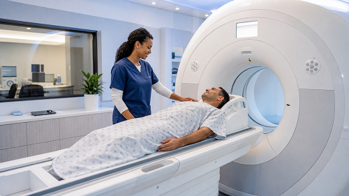
- You will lie on a table that will move into the scanner. The technologist will position you for the area being examined, and a coil that receives the signal may be placed around or over the area. If your head is scanned, the coil fits over your face without touching you, leaving space for you to see.
- Earplugs or headphones are required because MRI scanners are noisy; therefore, you need hearing protection. Hold the buzzer during the scan; press it anytime, and the radiographer can talk to you, stop the scan, or bring you out[4,5].
Keeping still
Staying still is necessary. Movement blurs MRI images, similar to photographs. Each sequence takes two to eight minutes, and you must remain still. Between sequences, you can swallow, blink, and move if the radiographer allows you to do so. For chest and abdominal scans, you may need to hold your breath for 15 to 20 s, and the radiographer will instruct you.
How long it takes
Scans last 20 to 60 minutes. A joint scan takes 20 to 30 minutes, a brain scan 30 to 45 minutes, and multi-region or contrast-enhanced scans take longer. Inquire when booking.
What it feels like
Nothing. MRI is painless. You may notice mild warmth in the scanned area; this is normal and is due to radio waves depositing energy in the tissue. Inform the radiographer if any spot feels hot, especially near a tattoo or implant. They will monitor you closely throughout.
Afterwards
Unless sedated, you can dress and leave the hospital immediately. There are no after-effects, recovery period, or restrictions on driving, work, eating, or exercise[4,5].
What is a contrast dye, and when is it used?
Not every MRI scan needs contrast. Many conditions can be seen clearly on an MRI without it. Your doctor recommends contrast only when it can provide extra information that may help with diagnosis or treatment.MRI contrast usually contains gadolinium and is injected into a vein. It changes the appearance of certain tissues on the scan, making some areas easier to see and compare with surrounding tissue.Contrast may be useful when doctors need to:
- Study a tumour more closely, for example, to see its size, borders, blood supply, or whether it has spread.
- Look for active inflammation or infection because affected areas may take up more contrast and become easier to identify.
- Examine blood vessels because contrast can make some blood vessels and abnormalities easier to see.
- Tell active disease from older damage,for example, in some brain conditions, contrast may help distinguish a new or active lesion from an older scar.
- Get more information about an abnormality already seen on a non-contrast scan.
Because contrast requires an additional medicine, an intravenous injection and additional imaging, it may increase the cost of the examination. It is therefore normally used when the extra information is expected to be useful for your care [5,6,7].
Understanding MRI Scan results
Who reads your scan and when
The radiographer who performs your scan will not interpret it. Your images are sent to a radiologist who reads the medical images. They look at the images, compare them with past scans, and write a report for the doctor who requested the scan. The process usually takes a few days to two weeks, but urgent scans are reported much faster, sometimes within an hour of the scan. Radiographers cannot tell you the results; if they are silent at the end, it is because of their professional limits with patients.
What the report will look like
Radiology reports have a standard format.
- Clinical indication: why the scan was requested
- Technique: which sequences were used and if contrast was given
- Findings: a detailed, neutral description of what is seen
- Impression or Conclusion: The radiologist's interpretation, which is the most important part.
If you receive a copy, read the impression first.
Words you may come across
Radiology language can sound frightening when it is simply accurate.
- Unremarkable/normal/no acute abnormality means nothing worrying is seen
- A lesion can be a cyst, scar, area of inflammation, or a tumour.
- Enhancement means the tissue took up contrast
- Signal change /hyperintense/ hypointense means the tissue appears brighter or darker than expected
- Oedema means swelling or excess fluid
- Degenerative changes means normal age-related wear, extremely common in the spine and joints
- Incidental finding means something found that has nothing to do with why you were scanned
- Correlate clinically means the radiologist is telling your doctor to interpret the image alongside your symptoms and examination
2 things you should know
- Incidental findings are common. MRI is very detailed and often detects harmless variations such as small cysts, benign lumps, and minor age-related changes. Most patients only require reassurance. Some patients require only one follow-up scan to confirm their stability.
- Imaging results do not always match the symptoms. Some people have disc bulges and degenerative changes on scans but no back pain.
Conversely, you may have pain but a normal MRI. However, a normal MRI doesn't mean your pain isn't real; it just means the cause isn't visible on an MRI. Both scenarios are common, which is why your doctor considers the report along with your personal situation.
Getting your results
When you have your scan, ask how and when you will receive the results of the scan. If possible, schedule a follow-up appointment before leaving. Write down your questions in advance, as appointments can be quick, and it is easy to forget important questions.
Benefits of an MRI Scan
MRI provides a safe and non-invasive method to view detailed images of the body's internal structures, allowing for more accurate medical evaluations and personalised treatment plans. The benefits of MRI are as follows:
No ionising radiation
MRI uses magnets and radio waves instead of X-rays. This is especially important for people who require frequent scans, children, pregnant women and imaging organs sensitive to radiation.
Exceptional soft tissue detail
MRI offers better soft tissue contrast than CT. It shows the brain, spinal cord, cartilage, ligaments, tendons, muscles, and internal structures of organs in ways that other tests cannot.
It can change what happens next for the better
The detailed information from an MRI can help doctors decide what treatment or further tests are needed. For example, MRI can help doctors target suspicious areas for prostate biopsy or detect some strokes that may be difficult to see on CT.
Any angle, any plane
MRI can create images in any direction without moving the patient and can be adjusted to highlight different tissue features, such as fluid, fat, blood flow, and water movement, in the same scan.
Non-invasive and painless
No cuts are made, no tools are involved, and no recovery time is needed.
This can reduce the need for more invasive tests.
An accurate MRI can sometimes avoid exploratory procedures or biopsies.
Risks of an MRI Scan
MRI is one of the safest imaging tests available. However, these risks are real and worth understanding.
Projectile injury
The magnet remains active even when no scanning is taking place. Any magnetic item brought into the room can be rapidly attracted to the magnet. As a result, access to the scanner room is strictly regulated, and the staff check everyone who enters the room.
Implants
Some devices can move, heat up, or malfunction in magnetic fields. Devices are classified as
- MR safe,
- MR conditional (safe under certain conditions)
- MR unsafe,
and radiographers check each device before scanning.
Metal in the eye
A metal fragment in or near the eye may move or heat up in the MRI scanner and could injure the eye. Tell the MRI staff if you have ever had a metal fragment enter your eye, particularly during welding, grinding or metal work. If there is any doubt that a fragment remains, the radiology team may arrange an X-ray or review a previous CT scan before allowing you to have the MRI.
Heating and burns
Radiofrequency energy heats the tissue, and although radiographers automatically control this, burns are still the most common MRI-related issue. These burns often occur when cables touch the skin, when the skin forms conductive loops, or when metallic objects are on the body.
Hearing
Scanner noise can be loud enough to harm your hearing if you do not use protection. You can prevent this by using the earplugs or headphones provided. Wear them correctly and ask for help if they don't fit well.
Claustrophobia and anxiety
Claustrophobia can cause scans to be stopped early. The strategies mentioned earlier aim to prevent this from happening. Addressing it early usually makes it manageable.
Sedation and anaesthesia
For young children or adults who cannot stay still or handle the scanner, doctors may use sedation or general anaesthesia to facilitate the procedure. This has small risks related to breathing and blood pressure and needs monitoring.
Pregnancy
MRI without contrast is safe for the foetus, and a study of over 1.4 million pregnancies found no increased risk of problems from first-trimester scans. Avoid gadolinium contrast during pregnancy unless absolutely necessary. Inform the radiographer if you are pregnant.
MRI and CT Scan. What is the Difference?
MRI CT
Conclusion
An MRI scan is a painless test that provides detailed pictures of the inside of your body without using ionising radiation. Before the scan, tell the radiology team about any implants, metal in your body, pregnancy or concerns about enclosed spaces. During the scan, wear the hearing protection provided, keep still and use the call buzzer if you become uncomfortable. Remember that an MRI image is only one part of your diagnosis. Your doctor will interpret the results together with your symptoms, examination and medical history.
Frequently Asked Questions (FAQs)
Does an MRI hurt?
No. You may feel mild warmth in the scanned area, and lying still may be uncomfortable; however, the scan itself is painless.
Can I have an MRI if I have a pacemaker or other implant?
Possibly. Many modern devices are MR Conditional, meaning MRI can be performed under specific conditions. The radiology team must check the exact device before the scan.
How long does an MRI take?
Most MRI scans take about 20–60 minutes, depending on the body area being examined and whether contrast is needed.
What if I panic inside the scanner?
Use the call buzzer. The radiographer can speak to you and stop the scan if necessary. Tell the department beforehand if you are worried about enclosed spaces.
Is MRI contrast dangerous?
For most people, gadolinium-based contrast agents are well tolerated. Tell your doctor or radiology team if you have kidney disease, have previously reacted to MRI contrast, or may be pregnant.
References
- Chalela JA, Kidwell CS, Nentwich LM, et al. Magnetic resonance imaging and computed tomography in emergency assessment of patients with suspected acute stroke: a prospective comparison. The Lancet 2007; 369: 293–298. Available from: https://pubmed.ncbi.nlm.nih.gov/17258669/
- Nguyen JC, Yaya-Quezada C, Lerebo WT, et al. MRI Diagnosis of Meniscus Tears in the Knee: An Updated Systematic Review and Meta-analysis. Radiology 2026; 319: e252288. Available from: https://pubmed.ncbi.nlm.nih.gov/42048590/
- Pedrosa I, Altman DA, Dillman JR, et al. American College of Radiology Manual on MR Safety: 2024 Update and Revisions. Radiology 2025; 315: e241405. Available from: https://pubs.rsna.org/doi/10.1148/radiol.241405
- US Food and Drug Administration. MRI (magnetic resonance imaging): benefits and risks. Silver Spring (MD): US Food and Drug Administration. Available from: https://www.fda.gov/radiation-emitting-products/mri-magnetic-resonance-imaging/benefits-and-risks:
- Ahn YH, Kang DY, Park S-B, et al. Allergic-like Hypersensitivity Reactions to Gadolinium-based Contrast Agents: An 8-year Cohort Study of 154 539 Patients. Radiology 2022; 303: 329–336. Available from: https://pubmed.ncbi.nlm.nih.gov/35191737/
- Woolen SA, Shankar PR, Gagnier JJ, et al. Risk of Nephrogenic Systemic Fibrosis in Patients With Stage 4 or 5 Chronic Kidney Disease Receiving a Group II Gadolinium-Based Contrast Agent: A Systematic Review and Meta-analysis. JAMA Intern Med 2020; 180: 223. Available from: https://pubmed.ncbi.nlm.nih.gov/31816007/
- Ray JG, Vermeulen MJ, Bharatha A, et al. Association Between MRI Exposure During Pregnancy and Fetal and Childhood Outcomes. JAMA 2016; 316: 952. Available from: https://jamanetwork.com/journals/jama/fullarticle/2547756
- Kasivisvanathan V, Rannikko AS, Borghi M, et al. MRI-Targeted or Standard Biopsy for Prostate-Cancer Diagnosis. N Engl J Med 2018; 378: 1767–1777. Available from: https://pubmed.ncbi.nlm.nih.gov/29552975/
Disclaimer:
The information provided on this website is for general educational and informational purposes only. It is not intended to replace professional medical advice, diagnosis, or treatment. Always seek the advice of your physician or other qualified health provider with any questions you may have regarding a medical condition.
Published April 25, 2026